Customer Spotlight- Ms. Kristie Huda- Tulane University
Kristie Huda is a Graduate Research Assistant and Ph.D. student at Tulane University. Ms. Huda has a background in Electrical Engineering and her Ph.D. work centers around Biomedical Engineering. Kristie is passionate about developing biomedical applications that will improve women’s health. Her research focuses on developing new imaging methods to study the dynamics of physiological function and molecular information. Kristie is currently working on developing methods to translate the photoacoustic system for human use in clinical settings. She utilizes both simulation and experimental work to develop and improve current imaging techniques.
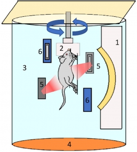
Abnormal placental function underlies many problems during pregnancy. Preeclampsia, gestational diabetes and fetal growth restriction can be examined by evaluating functional changes in the placenta. The challenge for medical science is evaluating this function in vivo, in real-time, and without harming the fetus. Ms. Huda and her colleagues recently published a paper titled “Spherical-view photoacoustic tomography for monitoring in vivo placental function” in which they examined a prototype photoacoustic tomography (PAT) system to measure oxygen saturation and folate kinetics in living mice placentas.
Photoacoustic systems provide higher contrast images of tissue function and composition than ultrasound alone. Photoacoustic tomography works by:
- A nanosecond laser is used to produce a pulse of coherent light which irradiates tissue
- The chromophores in the tissue absorb the light energy and convert it into heat energy resulting in expansion
- This expansion generates an ultrasonic (sound) wave which can be measured by an ultrasound transducer and processed to form an image
The lab’s PAT system was able to acquire a 3D volume scan that was fast and highly sensitive. Their work demonstrates that both physiological and molecular dynamics can be examined in vivo, in live placentas in real-time. These results have implications for diagnosing and treating problems during pregnancy as well as broader applications in cancer and other challenges to human health.
Q&A with Ms. Huda:
How did come to focus on women’s health and pursue a Ph.D. in Biomedical Engineering?
As a woman in science, I always wanted to contribute to the improvement of health care available for women. In my master’s in Biomedical Physics, I worked on a simulation project to identify malignancy of breast tumors using the Focused Impedance Method developed by my department at the University of Dhaka. During this process, I learnt how the progression of disease changes tissue properties and how to identify these changes noninvasively. This intrigued me so much that I decided to pursue a Ph.D. in biomedical engineering. While searching for positions in different research labs, my focus was on medical imaging and women’s health. Dr. Carolyn Bayer’s lab provides me the opportunity to develop methods for new imaging modalities for women.
What is still surprising to you in this field?
In this field, I am surprised by the constant innovation of new laser technology and computation power, which increase the potential of photoacoustic imaging to help us understand the physiopathology of diseases.
How does your OPOTEK Phocus HE benchtop enable your group’s work?
Our lab uses the OPOTEK Phocus HE benchtop laser for generating high contrast photoacoustic images. Using the high energy nanosecond pulsed laser, we study the change of oxygenation in abnormal placental tissue. It also enables us to understand the hemodynamics of the placenta using different contrast agents with the photoacoustic imaging modality.