Customer Spotlight- Dr. Raiyan Zaman, Harvard Medical School
Dr. Raiyan Zaman is an Assistant Professor in the Department of Radiology at Harvard Medical School and an Assistant Investigator at the Gordon Center for Medical Imaging at Massachusetts General Hospital. She did her Ph.D. work at the University of Texas at Austin where her focus was Biomedical Engineering Instrumentation. Dr. Zaman also did NIH T32 and American Heart Association Postdoctoral Fellowships at Stanford University School of Medicine in Cardiovascular Imaging before becoming an Instructor of Cardiovascular Medicine. For more information about Dr. Zaman and her group, please visit the lab’s website.
Coronary artery disease (CAD) is a leading cause of death worldwide. It is often caused by atherosclerosis- a progressive inflammatory condition in which deposits of plaque build up in the arteries of the heart, often resulting in heart attack. Early detection of these plaques is difficult due to their motion and size and because of the obscuring electrical signals of the heart. Identifying problem lesions that are likely to rupture would improve medical outcomes. Dr. Raiyan Zaman, in the Department of Radiology at Harvard Medical School, came up with a novel method to image these plaques using tunable laser light while she was a Post-doctoral Fellow at the Stanford University School of Medicine. Circumferential-Intravascular-Radioluminescence-Photoacoustic-Imaging (CIRPI) combines radioluminescence imaging and photoacoustic tomography with a new optical probe to achieve up to 63 times more signal to noise. In addition, photoacoustic imaging allows for the analysis of plaque composition and its morphology. This increase in sensitivity combined with information on the composition of the lesions is an exciting development in radiology and medicine. Dr. Zaman and her team are currently in the process of testing their system in an atherosclerotic animal model followed by clinical translation studies.

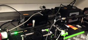
CIRPI system (picture courtesy of R. Zaman)
Q&A with Dr. Zaman:
How did you become interested in imaging?
During my graduate study, I was fascinated by optics and its interaction with human tissue. This inspired me to start designing novel optical imaging systems. However, when I moved to Stanford University School of Medicine for my T32 Postdoctoral Fellowship, I wanted to combine optical imaging systems with other imaging modalities such as photoacoustic and radioluminescence to overcome the limitation of optics in imaging. Since then my research has been focused on the design and development of catheter-based hybrid imaging for coronary artery disease.
How has your OPOTEK tunable laser system been useful in your lab?
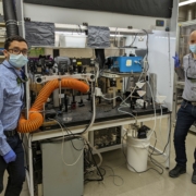
The OPOTEK laser is a key component of our CIRPI system for photoacoustic imaging. This small but powerful tunable laser is perfect for our portable imaging system; enabling us to wheel it to a patient’s bedside.
What outcome would you like to see as a result of your group’s work?
We are trying to minimize the multiple intravascular imaging procedures necessary for a patient who requires intervention for coronary artery disease. These imaging procedures are needed for the detection of stenosis and evaluation of coronary arterial wall before intervention. Our CIRPI system will combine all these necessary procedures into one imaging session.


